Stomach
The stomach is the expanded part of the digestive tract and receives the bouls from the oesophagus. In the stomach, the food is mixed and broken down by the stomach’s gastric secretions to form chyme. The stomach is subdivided anatomically into four regions.
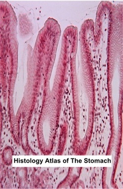
Histologically, the stomach only has three regions: the cardiac, fundic and pyloric regions. Each region possesses its own gastric mucosal glands. a. Gastric Mucosa The mucosa of the stomach has three specific structures. These structures include the following.
The gastric mucosa has three regions:
b. Gastric Submucosa The submucosa of the stomach is composed of dense connective tissue containing adipose tissue, blood vessels and Meissner’s submucosal plexus. The Meissner’s plexus innervates the vessels of the submucosa and the smooth muscle of the muscularis mucosae. c. Muscularis Externa The muscularis externa of the stomach is composed of three smooth muscle layers: an inner oblique layer, middle circular layer and outer longitudinal layer. The middle circular layer forms the pyloric sphincter. In addition, Auerbach’s myenteric plexus is located between the circular and longitudinal layers. d. Serosa The stomach is covered by a serosa, which is a connective tissue coat enveloped in the visceral peritoneum. Glands of the Gastric Mucosaa.Fundic Gastic Mucosa
The gastric mucosa of the fundic region presents fundic glands (gastric glands). These glands are simple, branched, tubular glands extending from the bottom of the gastric pits to the muscularis mucosa. The fundic glands are taller than the gastric pits. The gastric gland has three segments.
The fundic glands are composed of five cell types producing the gastric juice of the stomach (about 2L/day). The fundic region is the only one possessing all five cell types of gastric glands. These cells include the following. b.Cardiac Gastric Mucosa
Cardiac glands are found in the cardiac region, which surrounds the oesophageal orifice. Secretions of both the cardiac glands and the oesophageal cardiac glands contribute to the gastric juice and help protect the oesophageal epithelium against gastric reflux. The cardiac glands are occasionally branched, tubular glands, which are mainly composed of mucus-secreting cells with occasional interspersed enteroendocrine cells. In the cardiac glands, the gastric pits and glands have relatively the same length. The cardiac region glands have no chief cells and only a few parietal cells. The mucus-secreting cells have a flattened basal nucleus and mucin granules present in the apical cytoplasm. A short duct, composed of columnar cells with elongated nuclei, delivers mucous secretion into the lumen of the gastric pit. c.Pyloric Gastric Mucosa
The pyloric glands are located in the pyloric antrum between the fundus and the pylorus. They are branched, coiled, tubular glands that have relatively wide lumen. In the pyloric glands, the gastric pits are taller than the glands. Pyloric glands are composed of secretory cells that are similar in appearance to the surface mucous cells, which contain mostly mucous neck cells. They have no chief cells and only a few parietal cells and enteroendocrine cells are found interspersed within the gland epithelium. The pyloric glands’ secretion helps protect the pyloric mucosa. |
 (Alraddadi, 2011) |
Histology Text: Oesophagus - Small Intestine - Appendix - Large Intestine - Rectum & Anal Canal
Histology Atlas - Histopathology - Histology Quiz
Histology Atlas - Histopathology - Histology Quiz
