Liver
The liver is the largest gland in the body, weighing approximately 1,500 g and accounting for nearly 2.5% of the adult body weight. It has developed as an out-pouching from the wall of the foregut. It connects to the alimentary tract via the common bile duct, which opens in the duodenum.
Blood supply of the liver The liver is supplied by the hepatic artery and hepatic portal vein. The hepatic portal vein is the major source of the blood supply to the liver (about 75%). It carries venous blood that is largely depleted of oxygen from the digestive tract, pancreas and spleen. The portal vein blood contains the following.
Liver Functions
The liver has exocrine and endocrine functions:
Liver Capsule
A capsule of fibrous connective tissue called Glisson’s capsule invests the liver. A serous covering (visceral peritoneum) surrounds the capsule except where the liver adheres directly to the diaphragm or the other organs. Glisson’s capsule sends septa of connective tissue, stroma, into the substance of the liver subdivided the parenchyma into lobules. The place where the septa meet each other is called the portal area (portal triad). The portal areas contain bile ducts, lymph vessels and terminal branches of the hepatic artery and the portal veins. There is a small space at the edge of the portal canal, between the connective tissue stroma and the hepatocytes, called the space of Mall. It is filled with a small amount of connective tissue in which lymph drainage begins by means of blind-ended lymphatic capillaries. Parenchyma of the liver
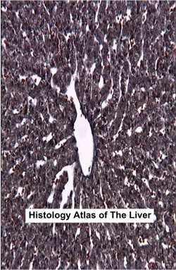
The parenchyma of the liver is composed of cells known as hepatocytes. They are organized into plates and separated by sinusoidal capillaries (sinusoids). Branches of the portal vein and hepatic artery found in the portal area supply these sinusoids. The sinusoids bathe the hepatocyte plates by carrying blood from the branches of the portal vein and hepatic artery found in the portal area to the central vein. Then blood drains to the sublobular veins, which in turn empty into the hepatic veins, which empty into the inferior vena cava. In addition to hepatocytes, the liver contains Kupffer cells (stellate sinusoidal macrophages) and Ito cells (hepatic stellate cells). Kupffer cells are macrophage cells derived from monocytes. They phagocyte foreign particulate matter and remove defunct red blood cells from the bloodstream. Ito cells are fat-storing cells that accumulate and store hepatic vitamin A. In pathological conditions, such as chronic inflammation or cirrhosis, Ito cells differentiate into myofibroblasts and synthesis collagen. Liver Lobules
Liver lobules are classified into three lobules: classic lobule, portal lobule and acinus of Rappaport. 1. Classic lobule The classic lobule is a hexagonal-shaped lobule presenting six portal areas (triads) at the apices and central vein at the centre of the lobule. Each classic lobule is composed of the anastomosed plates of the liver cells (hepatocytes). Sinusoids present between these plates and are lined with sinusoidal lining cells (endothelial cells), Kupffer cells and Ito cells. The space between the basal surface of hepatocytes and the basal surface of the sinusoid lining cells (endothelial and Kupffer cells) is known as the perisinusoidal space (space of Disse). Exchange materials between blood and liver cells occur in this space. Thus the hepatocytes do not come into contact with sinusoids. Intercellular spaces between liver cells are known as bile canaliculi. Bile passes peripherally from the bile canaliculi, which join to form the short intrahepatic ductules (canals of Hering). Intrahepatic bile ductules carry bile through the boundary of the lobule to the interlobular bile ducts at the portal areas, which lead to the common hepatic duct. 2. Portal lobule The portal lobule is a triangular-shaped lobule presenting three central veins at the apices and portal areas in the centre of the lobule. This organization is based on bile flow, the exocrine function of the liver, in which bile drains to the axial bile duct. 3. Acinus of Rappaport (liver/portal acinus) The acinus of Rappaport is a diamond-shaped lobule presenting two neighbouring central veins at the ends of the long axis and portal areas at the ends of the short axis. This organization is based on blood flow, where the terminal branches of the hepatic artery and portal vein supply the liver tissue. According to the distribution of oxygen and nutrients to the liver tissue, the hepatocytes in each liver acinus are arranged in three zones surrounding the short axis.
Hepatocytes
Hepatocytes are large, polygonal cells constituting about 80% of the cell population of the liver. They have large spherical nuclei occupying the centre of the cells. In the adult liver, many hepatocytes are binucleate. They have a long lifespan extending to five months. Liver cells are also capable of regenerating if they are lost to hepatotoxic processes, disease or surgery. In H&E staining, the hepatocyte has an acidophilic appearance. The cytoplasm contains rER, free ribosomes, numerous mitochondria, the Golgi apparatus, large numbers of peroxisomes, deposits of glycogen, lipid droplets and lysosomes. Peroxisomes are numerous in hepatocytes, and these are involved in many detoxification processes occurring in the liver, for example the detoxification of alcohol. The large Golgi apparatus in hepatocytes is concentrated near the bile canaliculus and is believed to be associated with the exocrine secretion of bile. Lysosomes dense bodies are seen in histologic sections concentrated near the bile canaliculus. They are the normal storage site for iron, which increases in a variety of pathologic conditions, for example hepatitis and anemia. |
 (Alraddadi, 2011) |
Histology Text: Major Salivary Glands - Gallbladder - Pancreas
Histology Atlas - Histopathology - Histology Quiz
Histology Atlas - Histopathology - Histology Quiz
